The Scan Is Not the Intervention
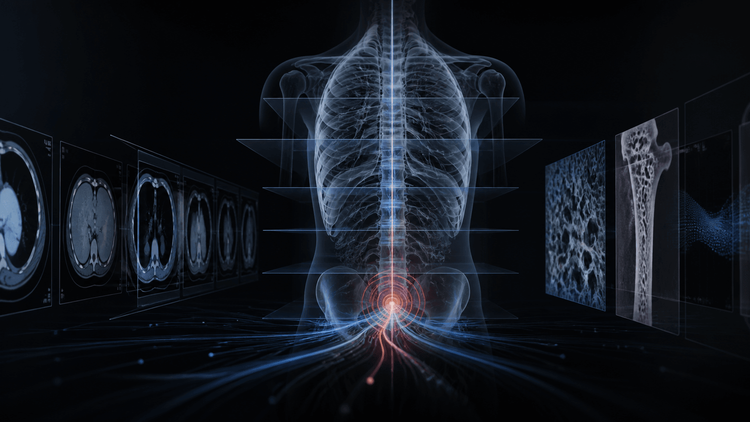
The next breakthrough in radiology AI will not be a model that sees more. It will be a system that makes sure the right next step actually happens.
For the last decade, the radiology AI conversation has been dominated by detection. Can the model find a pneumothorax? Can it mark a nodule? Can it quantify bone density? Can it identify a fracture, a bleed, a clot, or a suspicious lesion faster than a human reader?
Those are important questions. They are also incomplete.
A finding is not care. A signal is not an intervention. A report is not a pathway.
Radiology has always had this quiet paradox. A scan can reveal something clinically important, the report can be technically correct, and the patient can still receive no meaningful follow-up. An incidental vertebral compression fracture, coronary calcium, fatty liver, a suspicious pulmonary nodule, low bone density, or an early body-composition signal does not help a patient simply because it was visible on an image.
AI makes this paradox more urgent. It can search every eligible study, quantify signals consistently, and make overlooked information harder to miss. But it can also create a new pile of alerts with no owner, no documented hand-off, and no practical route to action.
Detection is a capability. Closure is the product.
The old framing stops too early
Most AI discussions in radiology still begin and end with model performance. Sensitivity. Specificity. AUC. Turnaround time. False positives. False negatives. Workflow impact. These metrics matter, and no serious clinical system can ignore them.
But they describe only the upstream part of the problem.
In real care, the question is not only whether the AI found the signal. The harder question is whether the signal survived the journey from image to report, from report to referrer, from referrer to patient, and from patient to a real clinical decision.
This is especially important for opportunistic imaging.
Opportunistic imaging means using studies already performed for one reason to identify clinically meaningful signals that may support prevention, risk stratification, or better care planning. A CT ordered for abdominal pain may contain information about bone density. A scan performed for oncology staging may carry body-composition information relevant to frailty, sarcopenia, surgical risk, treatment tolerance, and outcomes. A chest CT may show coronary calcium or emphysema that was not the original clinical question.
The opportunity is enormous because the data already exists.
But that is also the trap. Just because the signal is present does not mean the care system is ready to use it.
Opportunistic imaging is a pathway problem
The recent RSNA discussion of AI-assisted opportunistic osteoporosis screening is instructive. The model reviewed routine CT studies for low bone density. The technical achievement is important: hundreds of thousands of examinations, automated vertebral-body analysis, and strong radiologist agreement in sampled review.
But the more interesting part is what happens after detection.
At NYU Langone, a positive result does not simply become another isolated alert. It enters an EMR work list, and a dedicated bone-health team arranges DXA and referrals. That is the difference between an AI finding and a clinical program.
The model is only one part of the product. The pathway is the rest of the product.
That distinction should become central to how hospitals, radiologists, diagnostic networks, and AI companies think about the next phase of imaging AI. The real unit of innovation is not the algorithm alone. It is the operating model that turns an imaging signal into an accountable next step.
A useful opportunistic imaging program needs four verbs: detect, decide, route, and close.
The closed-loop scorecard
A mature radiology AI program should be judged by a cascade, not a single dashboard number.
Eligible studies. Which studies should the system evaluate, and which studies should be excluded because the indication, image quality, age group, prior history, or clinical context makes the signal inappropriate?
Valid signals. Which AI outputs meet a threshold that radiologists and clinicians believe is meaningful enough to communicate?
Communicated findings. Which findings are expressed in the report with clear, proportionate language that a referring doctor can act on?
Owned next steps. Which findings create a task for a real person or team: the referrer, a care navigator, a specialist clinic, a central clinical team, or the radiologist-led service layer?
Completed evaluation. Which patients actually complete the recommended next step, whether that is DXA, lab evaluation, specialist review, clinical counselling, treatment, or a documented decision not to proceed?
Action taken. Which cases result in a meaningful clinical action rather than another open loop?
This is the chart every opportunistic imaging program needs:
Eligible studies -> valid AI signals -> communicated findings -> owned next steps -> completed evaluation -> action taken
The early arrows measure technical performance and workflow reliability. The later arrows measure whether the program produces care.
An increase in detected findings with no increase in completed evaluation is not success. It is a queue.
The fragile step is routing
Every hospital can name an incidental finding. Fewer can say who owns it at 6 p.m. on a Friday.
That is where many AI deployments will fail. Not because the model is useless, but because the hand-off is weak.
Who receives the result? The reporting radiologist? The referring doctor? The ordering physician? A care navigator? A disease-specific clinic? A central operations team? What happens if the patient came from outside the system? What if the EMR is incomplete? What if there is no reliable phone number? What if the finding is real but not worth alarming the patient? What if the recommendation creates unnecessary downstream testing?
These are not administrative details. They are the clinical interface of the model.
Radiology AI is entering a stage where the hard work is no longer only detection. It is governance, language, thresholds, ownership, escalation, audit, and closure.
That is why radiologists matter more, not less.
The radiologist is not merely the person who checks whether the AI was right. The radiologist is the clinical authority who understands whether a signal should be reported, how uncertainty should be expressed, which recommendation is proportionate, and what would make the result useful to the clinician and patient.
In an AI-native radiology system, the radiologist moves from image interpreter to pathway architect.
India needs local pathway design
India should not import opportunistic imaging workflows as slide decks.
A high-volume diagnostic network, a tertiary hospital, a corporate hospital chain, a small referring ecosystem, and a distributed teleradiology network do not share the same source of truth. They do not have the same EMR maturity, follow-up capacity, patient contact workflows, clinician relationships, or tolerance for downstream investigation.
That means deployment has to be local.
Before scaling an opportunistic AI workflow, an Indian healthcare organization should answer five questions.
Who owns the output? A finding with no accountable recipient should not be counted as a clinical win.
What exact report language triggers action? The difference between "consider evaluation" and "recommend DXA/referral" is not cosmetic. It changes responsibility.
What happens when data is missing? Prior studies, clinical history, contact information, and EMR continuity are often incomplete. The fallback has to be designed.
How are false positives governed? A system that creates unnecessary anxiety, testing, or cost will lose trust even if the model performs well in a paper.
Which outcome is measured? Alerts generated, referrals accepted, evaluations completed, and treatment started are not the same metric.
This is where India can build something better than a copied workflow. The constraints are real: volume pressure, fragmented care, variable infrastructure, and shortage of specialists. But those constraints also force better service design.
The winning model will combine AI, radiologist judgment, structured reporting, central operations, and follow-up discipline into one clinical loop.
Sarcopenia shows where this is going
Sarcopenia and body-composition analysis are a powerful example of the next frontier.
Routine CT scans already contain information about muscle mass and body composition. In oncology, surgery, chronic disease, and elderly care, those signals can matter. They may help clinicians think about frailty, treatment tolerance, nutritional risk, rehabilitation, surgical planning, and outcomes.
But the same principle applies: the signal alone is not enough.
If a CT contains a body-composition signal suggestive of sarcopenia, what should happen next? Should it be reported for every patient or only selected populations? What threshold is clinically meaningful? Who receives the recommendation? Is there a nutrition pathway, geriatric pathway, oncology pathway, or pre-operative pathway ready to respond? How do we avoid overcalling, overburdening clinicians, or turning early risk signals into noise?
This is why the frontier is exciting.
The most important work is not just to identify more hidden signals in imaging. It is to turn those signals into disciplined clinical pathways that radiologists can trust, clinicians can use, and patients can benefit from.
That is the opportunity 5C is deeply interested in: not louder AI overlays, but better connections between image, interpretation, and accountable action.
A note to radiologists
The next decade of radiology will reward doctors who can do more than read fast.
Speed will still matter. Accuracy will still matter. Subspecialty depth will still matter. But AI will make another capability much more valuable: the ability to convert imaging knowledge into systems.
Radiologists will help define thresholds, reporting language, exclusions, escalation rules, audit cadence, and what counts as a closed loop. They will decide when an AI-generated signal is clinically meaningful, when it is premature, when it is unsafe, and when it should change the patient's next step.
That is a bigger role than report production.
It is clinical leadership inside an AI-native radiology system.
At 5C, we are building toward that future: radiology that is faster, more consistent, more specialized, and more connected to the care that follows the report. We are especially excited about frontier areas like opportunistic screening, body-composition intelligence, sarcopenia, quality systems, and AI-assisted reporting workflows that keep radiologists at the center of clinical accountability.
For radiologists who want to work at that frontier, the invitation is simple: help build the next radiology system with us.
Further reading
RSNA: AI Succeeds at Opportunistic Screening for Osteoporosis
Radiology: Deep Learning-based Opportunistic CT Osteoporosis Screening
RSNA: 2026 Knee Abnormality Detection AI Challenge
Radiology: Clinical History Summarization with Large Language Models
The scan is not the intervention. The intervention begins when a finding is owned, routed, and closed.